47+ Foot Bottom Diagram
Each foot pain diagram here is a useful visual tool for diagnosing what is wrong in your foot or ankle. In most two-footed and many four-footed animals the foot consists of.

Triple Speed Ethernet Megacore Function Guide Datasheet By Intel Digi Key Electronics
Web Understanding what is causing your foot pain is the first step to treating it.

. Bottom Plantar View of Foot. Web Foot in anatomy terminal part of the leg of a land vertebrate on which the creature stands. You can see the toes on.
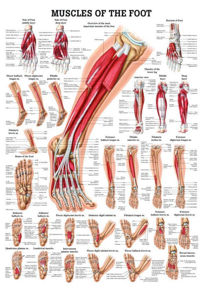
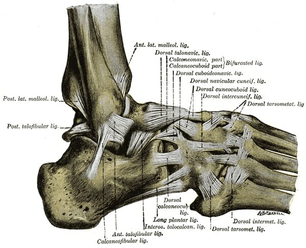
Web In fact there are so many ligaments that we need three different diagrams to show them all to you. They connect bones to other bones and are extremely important in stabilizing. Web The foots shape along with the bodys natural balance-keeping systems make humans capable of not only walking but also running climbing and countless.
It is made up of three joints. Web Match the corresponding numbers on the foot diagram below for a list of conditions that may be causing your foot and ankle pain. Available for both RF and RM licensing.
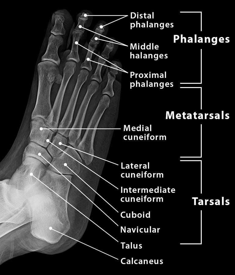
Web Columns of the Foot. Tarsals metatarsals and phalanges. Ligaments are strong connective tissue composed of fibrous tissues.
Ad Enjoy low prices on earths biggest selection of books electronics home apparel more. Web The foot is a complex anatomic structure composed of numerous bones joints ligaments muscles and tendons responsible for the complex coordinated. Web Match the corresponding numbers on the diagram below for a list of conditions that may be causing your foot and ankle pain.
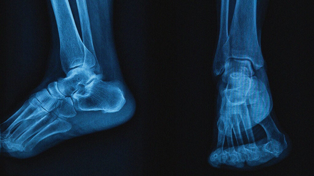
The ankle joint also known as the talocrural joint allows dorsiflexion and plantar flexion of the foot. Shop Alamy Prints Store - Crafted by Experts. This article provides a comprehensive Foot Pain Chart for all.
This diagram shows the sole of the foot. Web Ankle anatomy. The foot is sometimes described as having two columns Figure 3.
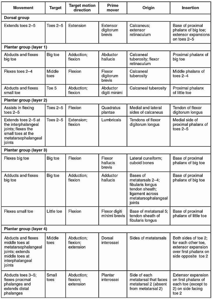
Web The 26 bones of the foot are divided into 3 groups. Web The bones of the foot are arranged to form 3 arches that give it the strength to support our bodies. Web The sural nerve branches from the tibial and common fibular nerves and is responsible for feeling on the outside of the foot and the small toe.
The medial column is more mobile and consists of the talus navicular medial. Acts as a lever for the. Web Find the perfect diagram of foot stock photo image vector illustration or 360 image.
The first two arches shape the bottom of the foot and the third. This is meant for educational purposes only. Web Foot pain can be debilitating and several conditions can cause pain in certain aspects of the foot.

Pin On Feet

Foot Charts
Foot Anatomy Pictures Models And Common Conditions Of The Foot
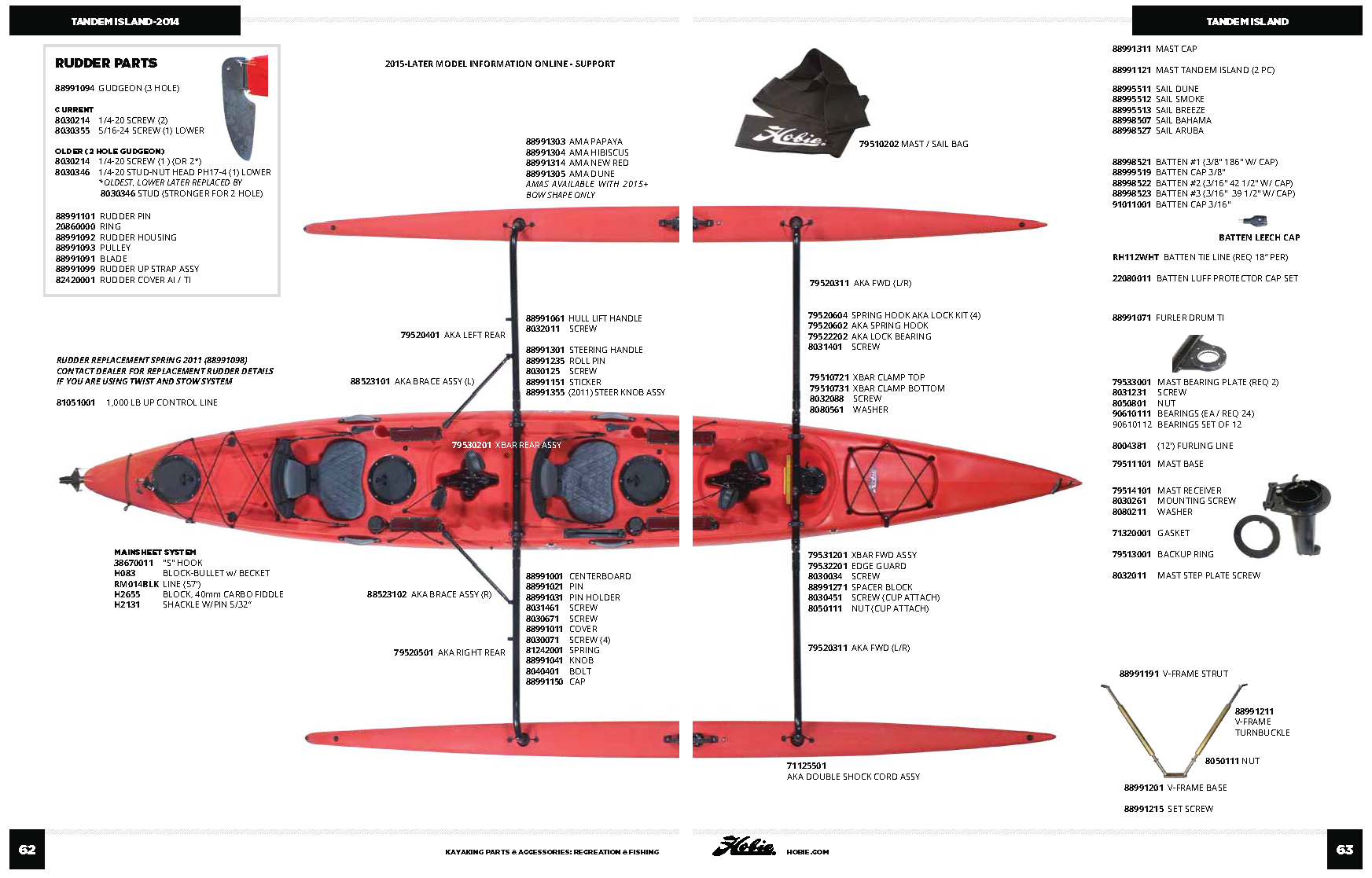
Hobie Tandem Island Parts Mariner Sails
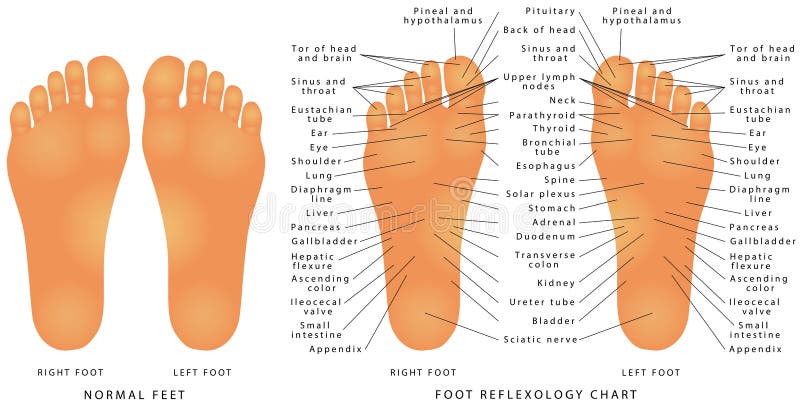
Reflexology Foot Chart Stock Illustrations 69 Reflexology Foot Chart Stock Illustrations Vectors Clipart Dreamstime
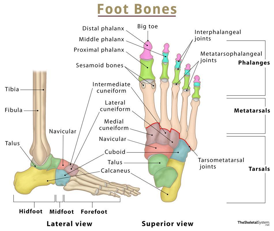
Foot Bones Names Anatomy Structure Labeled Diagrams
Foot Charts

Foot Anatomy Detail Picture Image On Medicinenet Com
Foot Bones Names Anatomy Structure Labeled Diagrams
Enhanced Collision Induced Unfolding And Electron Capture Dissociation Of Native Like Protein Ions Analytical Chemistry
Full Title For Class 305 Subclass 111
Foot Anatomy Bones Ligaments Muscles Tendons Arches And Skin

Foot Pain Chart Bottom Foot Pain Diagram
Boring Apparatus Black And White Stock Photos Images Alamy
Full Title For Class 305 Subclass 111

Reflexology Foot Chart Stock Illustrations 69 Reflexology Foot Chart Stock Illustrations Vectors Clipart Dreamstime
Foot Anatomy Bones Ligaments Muscles Tendons Arches And Skin